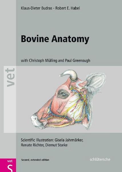
Produktbild: Bovine Anatomy

Bovine Anatomy An Illustrated Text

Aus der Reihe Schlütersche Vet

79,99 €

inkl. gesetzl. MwSt.

Beschreibung

Produktdetails

Format

PDF

Kopierschutz

Nein

Family Sharing

Ja

Text-to-Speech

Nein

Erscheinungsdatum

05.09.2011

Illustriert von

Gisela Jahrmärker + weitere

Herausgeber

Christoph K. W. Mülling + weitere

Verlag

Schlütersche eBooks

Seitenzahl

184 (Printausgabe)

Dateigröße

12893 KB

Auflage

2nd Enlarged edition (ENL).

Sprache

Deutsch, Englisch

EAN

9783842683594

Beschreibung

Zitat

...a 'must-have' book for anyone with an interest in bovine medicine... not only provides a detailed atlas of bovine anatomy; it also includes a comprehensive section on clinical anatomy... consistent layout of pages, with text on the left and drawings on the right... The large size is beneficial, the drawings are very clear and the illustration of structures from different angles makes a good visual aid for dissection; the colours are eye-catching and make it a pleasure just to leaf through the book... The coverage of material is comprehensive while remaining clinically relevant... The quality of the images and illustrations is superb and the descriptions of procedures are extremely helpful... very reasonable price --Renate Weller, Kate Holroyd, Andrea Turner and Peter Aitken, Veterinary Record, 22-Oct-2011

Produktdetails

Format

PDF

eBooks im PDF-Format haben eine festgelegte Seitengröße und eignen sich daher nur bedingt zum Lesen auf einem tolino eReader oder Smartphone. Für den vollen Lesegenuss empfehlen wir Ihnen bei PDF-eBooks die Verwendung eines Tablets oder Computers.

Kopierschutz

Nein

Dieses eBook können Sie uneingeschränkt auf allen Geräten der tolino Familie, allen sonstigen eReadern und am PC lesen. Das eBook ist nicht kopiergeschützt und kann ein personalisiertes Wasserzeichen enthalten. Weitere Hinweise zum Lesen von eBooks mit einem personalisierten Wasserzeichen finden Sie unter Hilfe/Downloads.

Family Sharing

Ja

Mit Family Sharing können Sie eBooks innerhalb Ihrer Familie (max. sechs Mitglieder im gleichen Haushalt) teilen. Sie entscheiden selbst, welches Buch Sie mit welchem Familienmitglied teilen möchten. Auch das parallele Lesen durch verschiedene Familienmitglieder ist durch Family Sharing möglich. Um eBooks zu teilen oder geteilt zu bekommen, muss jedes Familienmitglied ein Konto bei Thalia oder einem anderen tolino-Buchhändler haben. Weitere Informationen finden Sie unter Hilfe/Family-Sharing.

Text-to-Speech

Nein

Bedeutet Ihnen Stimme mehr als Text? Mit der Funktion Text-to-Speech können Sie sich im tolino webReader und in der aktuellen Thalia – Lesen & Hören App das eBook vorlesen lassen. Weitere Informationen finden Sie unter Hilfe/Text-to-Speech.

Barrierefreiheit

  • keine Information zur Barrierefreiheit bekannt

Erscheinungsdatum

05.09.2011

Illustriert von

  • Gisela Jahrmärker
  • Renate Richter
  • Diemut Starke

Herausgeber

Verlag

Schlütersche eBooks

Seitenzahl

184 (Printausgabe)

Dateigröße

12893 KB

Auflage

2nd Enlarged edition (ENL).

Sprache

Deutsch, Englisch

EAN

9783842683594

Noch keine Bewertungen vorhanden

Verfassen Sie die erste Bewertung zu diesem Artikel

Helfen Sie anderen Kundinnen und Kunden durch Ihre Meinung.

Kundinnen und Kunden meinen

Bewertungen (0)

Die Leseprobe wird geladen.
  • Produktbild: Bovine Anatomy
  • Preface Topographic Anatomy Thoracic limb (A. Wunsche, R. Habel and K.-D. Budras) Skeleton of the thoracic limb Muscles and nerves of the shoulder, arm, and forearm Cutaneous nerves, blood vessels, and lymph nodes of the thoracic limb Vessels and nerves of the manus Interdigital nerves and vessels, interossei, and fasciae of the manus Synovial structures of the thoracic limb Pelvic limb (A. Wunsche, R. Habel and K.-D. Budras) Skeleton of the pelvic limb Lateral thigh and cranial crural muscles with their nerves Medial thigh and caudal crural muscles with their nerves Cutaneous nerves, blood vessels, and lymph nodes of the pelvic limb Arteries, veins, and nerves of the pes Dermis of the hoof (Ch. Mulling and K.-D. Budras) The hoof (ungula) (Ch. Mulling and K.-D. Budras) Synovial structures of the pelvic limb (Ch. Mulling and K.-D. Budras) Head (R. Habel, and K.-D. Budras) Skull and hyoid apparatus (R. Habel and K.-D. Budras) Skull with teeth (R. Habel and K.-D. Budras) Skull with paranasal sinuses and horns (R. Habel and K.-D. Budras) Superficial veins of the head, facial n. (VII), and facial muscles (S. Buda and K.-D. Budras) Trigeminal n. (V3 and V2), masticatory mm., salivary gll., and lymphatic system (S. Buda and K.-D. Budras) Accessory organs of the eye (P. Simoens and K.-D. Budras) The eyeball (bulbus oculi) (P. Simoens and K.-D. Budras) Nose and nasal cavities, oral cavity and tongue (S. Buda, R. Habel, and K.-D. Budras) Pharynx and larynx (S. Buda, R. Habel and K.-D. Budras) Arteries of the head and head-neck junction, the cran. nn. of the vagus group (IX-XI), and the hypoglossal n. (XII)(S. Buda and K.-D. Budras) Central nervous system and cranial nerves The brain (R. Habel and K.-D. Budras Cranial nerves I-V (S. Buda, H. Bragulla and K.-D. Budras) Cranial nerves VI-XII (S. Buda, H. Bragulla, and K.-D. Budras) Spinal cord and autonomic nervous system (S. Buda and K.-D. Budras) Vertebral column, thoracic skeleton, and neck (A. Wunsche, R. Habel and K.-D. Budras) Vertebral column, ligamentum nuchae, ribs, and sternum Neck and cutaneous muscles Deep shoulder girdle muscles, viscera and conducting structures of the neck Thoracic cavity Respiratory muscles and thoracic cavity with lungs (Ch. Mulling and K.-D. Budras) Heart, blood vessels, and nerves of the thoracic cavity (R. Habel and K.-D. Budras) Abdominal wall and abdominal cavity The abdominal wall (R. Habel, A. Wunsche and K.-D. Budras) Topography and projection of the abdominal organs on the body wall Stomach with rumen, reticulum, omasum, and abomasum (A. Wunsche and K.-D. Budras) Blood supply and innervation of the stomach; lymph nodes and omenta (R. Habel, A. Wunsche and K.-D. Budras) Spleen, liver, pancreas, and lymph nodes (P. Simoens, R. Habel and K.-D. Budras) Intestines with blood vessels and lymph nodes (P. Simoens, R. Habel and K.-D. Budras) Pelvic cavity and inguinal region, including urinary and genital organs Pelvic girdle with the sacrosciatic lig. and superficial structures in the pubic and inguinal regions (R. Habel and K.-D. Budras) Inguinal region with inguinal canal, inguinal lig., and prepubic tendon (R. Habel and K.-D. Budras) Lymphatic system, adrenal glands, and urinary organs (K.-D. Budras and A. Wunsche) Arteries, veins, and nerves of the pelvic cavity (A. Wunsche and K.-D. Budras) Female genital organs (H. G. Liebich and K.-D. Budras) The udder (H. Bragulla, H. Konig, and K.-D. Budras) The udder with blood vessels, lymphatic system, nerves, and development (H. Bragulla, H. Konig, and K.-D. Budras) Male genital organs and scrotum (R. Habel and K.-D. Budras). Perineum, pelvic diaphragm, ischiorectal fossa, and tail (R. Habel and K.-D. Budras) Anatomical aspects of bovine spongiform encephalopathy (BSE) (S. Buda, K.-D. Budras, T. Eggers, R. Fries, R. Habel, G. Hildebrandt, K. Rauscher, and P. Simoens) Special Anatomy, Tabular Part Myology Lymphatic system Peripheral nervous system Contributions to Clinical-Functional Anatomy Applied anatomy of the carcass (K.-D. Budras, R. Fries, and R. Berg) References Index