















































KI-Beratung Beta

Gutscheinbedingungen

Gültig bis 11.11.2026 | Gültig für Spielzeug, Filme, Musik, Software, Games, Geschenke & Trends, Schreibwaren, Hörbücher und Hörbuch-Downloads (außer Abo & Shelfies), gebrauchte Bücher, nicht preisgebundene Bücher und Kalender | Einlösbar unter thalia.de und in der Thalia App | Click & Collect nur mit Online-Zahlung (Paypal/Kreditkarte) vorab | Einzelne Artikel können ausgeschlossen sein | Nicht kombinierbar mit anderen Gutscheinen oder Preisaktionen | Nur einmal pro Einkauf einlösbar | Gutschein wird auf max. 500€ Bestellwert angerechnet | Keine Barauszahlung | Digitale Hörbücher nur für Android  | Nicht gültig für preisgebundene Artikel (aufgrund der Buchpreisbindung sind Gutscheine nicht auf Bücher, eBooks, Presse einlösbar), tolino eReader & Zubehör, Abonnements & Flatrates, Geschenkkarten, Versandkosten und Services

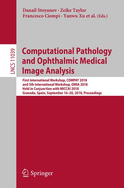
  • Produktbild: Computational Pathology and Ophthalmic Medical Image Analysis
  • Produktbild: Computational Pathology and Ophthalmic Medical Image Analysis
Band 11039

Computational Pathology and Ophthalmic Medical Image Analysis First International Workshop, COMPAY 2018, and 5th International Workshop, OMIA 2018, Held in Conjunction with MICCAI 2018, Granada, Spain, September 16 - 20, 2018, Proceedings

49,99 €

inkl. gesetzl. MwSt., Versandkostenfrei


Beschreibung

This book constitutes the refereed joint proceedings of the First International Workshop on Computational Pathology, COMPAY 2018, and the 5th International Workshop on Ophthalmic Medical Image Analysis, OMIA 2018, held in conjunction with the 21st International Conference on Medical Imaging and Computer-Assisted...

Beschreibung

This book constitutes the refereed joint proceedings of the First International Workshop on Computational Pathology, COMPAY 2018, and the 5th International Workshop on Ophthalmic Medical Image Analysis, OMIA 2018, held in conjunction with the 21st International Conference on Medical Imaging and Computer-Assisted Intervention, MICCAI 2018, in Granada, Spain, in September 2018.

The 19 full papers (out of 25 submissions) presented at COMPAY 2018 and the 21 full papers (out of 31 submissions) presented at OMIA 2018 were carefully reviewed and selected. The COMPAY papers focus on artificial intelligence and deep learning. The OMIA papers cover various topics in the field of ophthalmic image analysis.

Produktdetails

Einband

Taschenbuch

Erscheinungsdatum

14.09.2018

Abbildungen

XVII, 347 p. 135 illus.

Herausgeber Jeroen van der Laak + weitere

Produktdetails

Einband

Taschenbuch

Erscheinungsdatum

14.09.2018

Abbildungen

XVII, 347 p. 135 illus.

Herausgeber
Verlag Springer
Seitenzahl

347

Maße (L/B/H)

23,5/15,5/2 cm

Gewicht

557 g

Auflage

1st ed. 2018

Sprache

Englisch

ISBN

978-3-030-00948-9

Herstelleradresse

Springer-Verlag KG
Sachsenplatz 4-6
1201 Wien
AT

Email: ProductSafety@springernature.com

Ein neues Kapitel für Ihre Bücher

Ein neues Kapitel für Ihre Bücher

Schenken Sie Ihren alten Schätzen ein zweites Leben und erhalten Sie dafür eine Thalia Geschenkkarte.

Jetzt verkaufen
Jetzt verkaufen

Noch keine Bewertungen vorhanden

Verfassen Sie die erste Bewertung zu diesem Artikel

Helfen Sie anderen Kundinnen und Kunden durch Ihre Meinung.

Kundinnen und Kunden meinen

Bewertungen (0)

  • Produktbild: Computational Pathology and Ophthalmic Medical Image Analysis
  • Produktbild: Computational Pathology and Ophthalmic Medical Image Analysis
  • Improving Accuracy of Nuclei Segmentation by Reducing Histological Image Variability.- Multi-Resolution Networks for Semantic Segmentation in Whole Slide Images.- Improving High Resolution Histology Image Classification with Deep Spatial Fusion Network.- Construction of a Generative Model of H&E Stained Pathology Images of Pancreas Tumors Conditioned by a Voxel Value of MRI Image.- Accurate 3D reconstruction of a whole pancreatic cancer tumor from pathology images with different stains.- Role of Task Complexity and Training in Crowdsourced Image Annotation.- Capturing global spatial context for accurate cell classification in skin cancer histology.- Exploiting Multiple Color Representations to Improve Colon Cancer Detection in Whole Slide H&E Stains.- Leveraging Unlabeled Whole-Slide-Images for Mitosis Detection.- Evaluating Out-of-the-box Methods for the Classification of Hematopoietic Cells in Images of Stained Bone Marrow.- DeepCerv: Deep neural network for segmentation free robustcervical cell classification.- Whole slide image registration for the study of tumor heterogeneity.- Modality Conversion from Pathological Image to Ultrasonic Image Using Convolutional Neural Network.- Structure instance segmentation in renal tissue: a case study on tubular immune cell detection.- Cellular Community Detection for Tissue Phenotyping in Histology Images.- Automatic Detection of Tumor Budding in Colorectal Carcinoma with Deep Learning.- Significance of Hyperparameter Optimization for Metastasis Detection in Breast Histology Images.- Image Magnification Regression Using DenseNet for Exploiting Histopathology Open Access Content.- Uncertainty Driven Pooling Network for Microvessel Segmentation in Routine Histology Images.- Ocular Structures Segmentation from Multi-sequences MRI using 3D Unet with Fully Connected CRFs.- Classification of Findings with Localized Lesions in Fundoscopic Images using a Regionally Guided CNN.- Segmentation of Corneal Nerves Using a U-Net-based Convolutional Neural Network.- Automatic Pigmentation Grading of the Trabecular Meshwork in Gonioscopic Images.- Large Receptive Field Fully Convolutional Network for Semantic Segmentation of Retinal Vasculature in Fundus Images.- Explaining Convolutional Neural Networks for Area Estimation of Choroidal Neovascularization via Genetic Programming.- Joint Segmentation and Uncertainty Visualization of Retinal Layers in Optical Coherence Tomography Images using Bayesian Deep Learning.- cGAN-based lacquer cracks segmentation in ICGA image.- Localizing Optic Disc and Cup for Glaucoma Screening via Deep Object Detection Networks.- Fundus Image Quality-guided Diabetic Retinopathy Grading.- DeepDisc: Optic Disc Segmentation based on Atrous Convolution and Spatial Pyramid Pooling.- Large-scale Left and Right Eye Classification in Retinal Images.- Automatic Segmentation of Cortex and Nucleus in Anterior Segment OCT Images.- Local Estimation of the Degree of Optic Disc Swelling from Color Fundus Photography.- Visual Field based Automatic Diagnosis of Glaucoma Using Deep Convolutional Neural Network.- Towards standardization of retinal vascular measurements: on the effect of image centering.- Feasibility study of Subfoveal Choroidal Thickness Changes in Spectral-Domain Optical Coherence Tomography Measurements of Macular Telangiectasia Type 2.- Segmentation of retinal layers in OCT images of the mouse eye utilizing polarization contrast.- Glaucoma Diagnosis from Eye Fundus Images Based on Deep Morphometric Feature Estimation.- 2D Modeling and Correction of Fan-beam Scan Geometry in OCT.- A Bottom-up Saliency Estimation Approach for Neonatal Retinal Images.