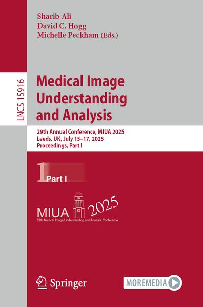
Produktbild: Medical Image Understanding and Analysis
Band 15916 - 10%

Medical Image Understanding and Analysis 29th Annual Conference, MIUA 2025, Leeds, UK, July 15–17, 2025, Proceedings, Part I

10% sparen

85,99 € UVP 96,29 €

inkl. gesetzl. MwSt., Versandkostenfrei


Beschreibung

Produktdetails

Einband

Taschenbuch

Erscheinungsdatum

17.07.2025

Abbildungen

XXI, 320 p. 106 illus., 94 illus. in color.

Herausgeber

Sharib Ali + weitere

Verlag

Springer

Seitenzahl

320

Maße (L/B/H)

23,5/15,5/1,9 cm

Gewicht

522 g

Sprache

Englisch

ISBN

978-3-031-98687-1

Beschreibung

Produktdetails

Einband

Taschenbuch

Erscheinungsdatum

17.07.2025

Abbildungen

XXI, 320 p. 106 illus., 94 illus. in color.

Herausgeber

Verlag

Springer

Seitenzahl

320

Maße (L/B/H)

23,5/15,5/1,9 cm

Gewicht

522 g

Sprache

Englisch

ISBN

978-3-031-98687-1

Herstelleradresse

Springer-Verlag KG
Sachsenplatz 4-6
1201 Wien
AT

Email: ProductSafety@springernature.com

Ein neues Kapitel für Ihre Bücher

Ein neues Kapitel für Ihre Bücher

Schenken Sie Ihren alten Schätzen ein zweites Leben und erhalten dafür eine Thalia Geschenkkarte.

Jetzt verkaufen
Jetzt verkaufen

Noch keine Bewertungen vorhanden

Verfassen Sie die erste Bewertung zu diesem Artikel

Helfen Sie anderen Kundinnen und Kunden durch Ihre Meinung.

Kundinnen und Kunden meinen

Bewertungen (0)

  • Produktbild: Medical Image Understanding and Analysis

  • .-
    Frontiers in Computational Pathology
    .


    .- Transductive Survival Ranking for Pan-cancer Automatic Risk Stratification using Whole Slide Images.


    .- Benchmarking Histopathology Foundation Models in a Multi-center Dataset for Skin Cancer Subtyping.


    .- MitoNet: Efficient Ki-67 Detection in H\&E-Stained Images.


    .- ASTER: Automated Segmentation of Endometrial Histology Images for Reproductive Health Assessment.


    .- Leveraging Pathology Foundation Models for Panoptic Segmentation of Melanoma in H\&E Images.


    .- SMatt-DINO: Spatially Aware Masked Attention Network for High Resolution Brain Image Classification.


    .- Persistent Homology and Gabor Features Reveal Inconsistencies Between Widely Used Colorectal Cancer Training and Testing Datasets.


    .- SWIFT-Reg: Slide-Wide Intelligent Feature-based Tissue Registration.


    .- Learnable Moran’s Index for Modeling Spatial Autocorrelation in Whole Slide Images to Predict Breast Cancer Outcomes.


    .-
    Image Synthesis and Generative Artificial Intelligence
    .


    .- Augmenting Chest X-ray Datasets with Non-Expert Annotations.


    .- Leveraging Synthetic Data for Whole-Body Segmentation in X-ray Images.


    .- Transform(AI)ng Radiology with CheXSBT: Integrating Dual-Attention Swin Transformer with BERT for Seamless Chest X-Ray Report Generation.


    .- Cardiac Ultrasound Video Generation Using a Diffusion Model with Temporal Transformer.


    .- KCLVA: Knowledge-enhanced Contrastive Learning and View-specific Attention for Chest X-ray Report Generation.


    .- BlastDiffusion: A Latent Diffusion Model for Generating Synthetic Embryo Images to Address Data Scarcity in In Vitro Fertilization.


    .- MediAug: Exploring Visual Augmentation in Medical Imaging.


    .- On the Robustness of Medical Vision-Language Models: Are they Truly Generalizable?.


    .- DiNO-Diffusion: Scaling Medical Diffusion Models via Self-Supervised Pre-Training.


    .- Knowledge-Driven Hypothesis Generation for Burn Diagnosis from Ultrasound with Vision-Language Model.


    .- Multimodal Federated Learning With Missing Modalities through Feature Imputation Network.


    .- Parameter-Efficient Multimodal Adaptation for Certified Robustness of Medical Vision-Language Models.